Palaeohistology
The following list is a compilation of histological articles that deal directly or indirectly with skeletochronology or life history data based on bone microstructures of fossil amniotes. We realise that this list of articles and the respective taxa studied, although comprehensively covering major works in the field of bone histology of fossil amniotes, is not exhaustive. Based on the scope of the project, we did not include works that deal with dental hard tissues, bone pathologies (Rothschild and Martin, 1993; see also annotated bibliography on dinosaur bone pathology by Tanke and Rothschild, 2002), or bone diagenesis. Similarly, bone histology on bones from archaeological sites is underrepresented. Further literature overviews, also on general aspects of bone histology, can be found for example in Ricqlčs (1977, 1980), Chinsamy-Turan (2005) and Francillon-Vieillot et al. (1990).
There are few studies (Hill, 2005; Scheyer, 2007a), which broadly sampled the amniote tree of life (including synapsids, parareptilians and eureptilians) for bone histology, for which we refrain from giving complete lists of studied taxa here.
Recommended Literature:
- Scheyer, T. M., N. Klein, and P. M. Sander. 2010. Developmental palaeontology of Reptilia as revealed by histological studies. In: Developmental Vertebrate Palaeontology (Sánchez-Villagra, M. R., ed.). Seminars in Cell and Developmental Biology [doi: 10.1016/j.semcdb.2009.11.005].

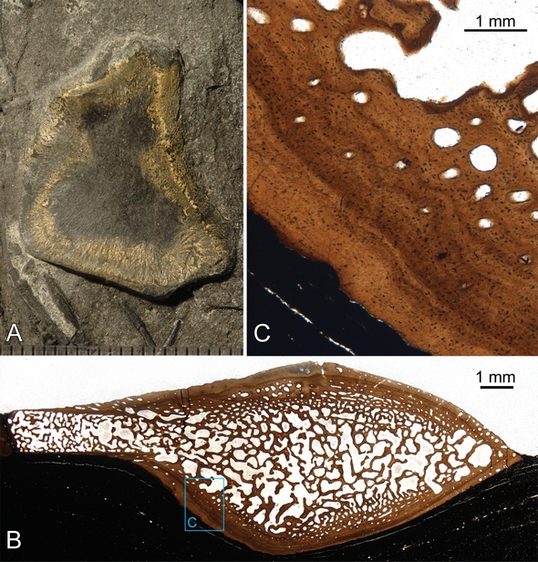
Outer morphology and bone histology of a humerus of the small ichthyosaur Mixosaurus from the Middle Triassic UNESCO world heritage site Monte San Giorgio, Switzerland. The specimen belongs to the collections of the Palaeontological Institute and Museum of the University of Zurich. A) Humerus preserved in the sediment prior to sampling. B) Complete thin-section of the specimen in normal transmitted light. Compact bone surrounds interior cancellous bone. The humerus grows by adding circumferential periosteal layers of bone on the outside, whereas the interior spongiose center of the bone enlarges as erosion cavities continuously erode the inner layers of the compact cortical bone. C) Detail of the outer compact bone showing a reduced amount of vascular canals, as well as growth marks (darker lines) in the bone tissue. The position of the close-up is marked by the inset rectangle in B). Picture copyright: Torsten Scheyer